A skin tag is a benign small skin growth, often attached by a thin stalk. At Experience Care on Montreal’s North Shore, our physician differentiates true skin tags from lesions requiring further assessment—unusual color, rapid growth, broad base, or ulceration are signs that warrant further evaluation.
.png)

.png)
.png)
A skin tag is often a small soft growth, sometimes attached by a thin base. It can appear in areas of friction, including the eyelids and the area around the eyes.
It is typically benign. However, near the eye, several lesions can look alike: milium grain, xanthhelasma, keratosis, wart, papilloma, or more suspicious lesions.
On the eyelid, distinguishing a skin tag from a molluscum, a viral wart, or an early carcinoma completely changes the approach.
THE CARE EXPERIENCE
Symptoms
A skin tag is often silent, but it can become bothersome when it catches, rubs, or is near the edge of the eyelid.
01
A small, soft growth, like a skin tag or a soft bump, attached to the eyelid or the skin around the eye.
02
The lesion can catch on makeup, glasses, during cleaning, or when blinking.
03
A skin tag can become tender or bleed slightly if it is irritated or twisted.
When the skin tag becomes visible and bothersome, especially on the eyelids where makeup catches on it.
Feeling of something rubbing or moving with each blink, especially along the eyelid margin.
A lesion that becomes darker, takes on multiple shades, or changes color. This is distinct from the typical skin color of a skin tag.
A lesion that grows slowly over months or years, without sudden changes.
Doubt about what it is exactly, especially if its appearance does not resemble a classic skin tag (soft, pedunculated, skin-colored).
Three questions guide the choice: is it a true skin tag, is the base thin (pedunculated) or broad, and does removal by destruction suffice or is analysis needed?
A true skin tag allows for several approaches. The right choice depends on the base of the lesion (thin or broad stalk), the distance from the lash line, and whether the diagnosis is certain or not.
Schedule an assessment"
Near the eye, the physician first assesses the appearance, evolution, and location of a small lesion before discussing removal.
Dr. Karen Dzolang, family physician
Overview
These guidelines do not replace a clinical examination. They illustrate why a typical skin tag is easily removed, while a lesion with a broad base or unusual appearance requires a different approach.
Key Indicators to Observe
Four situations, three possible approaches
Typical skin tag, irritated lesion, broad base, atypical appearance—each profile guides to a different approach: local removal, biopsy, or monitoring.
01
Typical
A typical skin tag is often soft, attached by a small base, and not very painful.
Signs
•
Soft or pliable appearance
•
Thin base or small stalk
•
Slow or stable progression
02
Irritated
An irritated or inflamed skin tag that has caught or twisted. Local removal reduces discomfort and limits recurrence.
Signs
•
Less narrow base
•
Border to examine
•
Healing expectations to discuss
03
Broad base
When the base is wide rather than pedunculated, the lesion may be something other than a simple skin tag (wart, viral papilloma, lesion to analyze). Excision with sampling is often preferable.
Signs
•
Redness after rubbing
•
Minor bleeding after irritation
•
Discomfort during cleaning or makeup
04
Atypical
Rapid growth, crusting, a wound that does not close, localized eyelash loss. These signs go beyond the typical presentation of a skin tag and suggest a biopsy or specialist consultation.
Signs
•
Crust or ulceration
•
Rapid change
•
Loss of eyelashes or distorted eyelid margin
To Check
01
A small growth can form in an area of friction or a skin fold, including near the eye.
02
Blinking, cleaning, makeup, or glasses can make the lesion more uncomfortable.
03
The doctor examines the appearance, then chooses between surveillance, local removal, or referral if necessary.
Origine
Skin tags are common and often associated with friction, skin folds, aging, family predisposition, and certain metabolic contexts.
Associations with diabetes, insulin resistance, or metabolic syndrome do not allow for a diagnosis based solely on a lesion. However, they may guide the discussion if there are many or new skin tags.
Perspective
A skin tag may remain stable for a long time or become bothersome if it catches, rubs, or bleeds. Removal depends on its appearance, location, proximity to the eye, and the limitations discussed during the consultation.
A typical skin tag does not necessarily need to be removed. It can remain stable for a long time and simply be monitored if it doesn’t change or cause discomfort.
However, it may become more bothersome if it snags, rubs, bleeds after irritation, or is located near the edge of the eyelid. At that point, the approach primarily depends on the base of the lesion (thin for simple removal, broad for excision) and its distance from the lash line.
An evaluation is useful if the lesion changes rapidly, bleeds without friction, ulcers, or alters the eyelid edge or eyelashes.
A lesion that grows visibly in a few weeks, whereas a skin tag usually develops over months or years.
Attention
Blood appearing without scratching the lesion, or bleeding that recurs with the slightest touch.
Attention
A scab that reappears after healing, or a sore that doesn't close — typical of early-stage skin cancers.
Attention
A very dark color, multiple shades, or changing borders — a skin tag is usually skin-colored.
Attention
Eyelashes falling out at the site of the lesion, or an eyelid margin that changes shape — the lesion damages the eyelash root.
Attention

Family doctor and graduate of the Université de Montréal, in good standing with the Collège des médecins du Québec (CMQ), Dr. Karen Dzolang serves as medical director for the CARE network. For eyelid lesions and skin plaques near the eye, she helps frame the evaluation, possible indications, treatment limits and situations where another medical opinion is preferable.
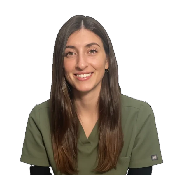
A registered nurse trained in pediatrics at Sainte-Justine Hospital, Karine Charbonneau later specialized in the dry eye clinic. Recognized by her patients for her gentleness, patience, and attentiveness, she supports each individual with precision and care, from the first appointment to long-term follow-up.

With an approach that combines Softness, Transparency and Great Meticulousness, Carolane ensures that each patient immediately feels confident and safe.
Its objective is simple: to make your care experience as comfortable as it is effective.
With a rich nursing experience that began in 2014, Carolane has enriched her expertise with a background in Medical Aesthetic Treatments To offer you the most recent protocols.
Passionate about improving the quality of life, she is entirely dedicated to supporting you with listening and professionalism throughout your career at the CARE clinic.