A milia looks like a small, firm white spot near the eye. At Experience Care, the doctor examines its color, texture, location, and changes before discussing removal options.
.png)
A milium typically appears as a small white or yellowish, firm, and smooth dot. It often appears on the face, cheeks, eyelids, or around the eyes.
It forms when keratin gets trapped beneath a thin layer of skin. There isn't always a single visible cause, and several eyelid lesions can look similar.
Near the eye, the issue is not just aesthetic: it’s important to avoid puncturing, scraping, or burning a lesion without knowing what it is.
THE CARE EXPERIENCE
We observe size, color, texture, and location before discussing removal.
Picking or scratching near the eye can irritate the skin, create a mark, or cause an infection.
A stable spot far from the eye can be monitored; an accessible spot might sometimes be removed; a changing, irritated lesion, or one near the eyelash line should be handled with more caution.
Symptômes
Milia are often silent. The number of dots, their location, pain, redness, bleeding, or lash loss indicate whether to simply monitor or consult promptly.
01
A small white or yellowish bump may remain under the skin without draining like a pimple.
02
Milia often appear on the eyelids, cheeks, or around the eyes.
03
A typical milium is often painless, without significant redness or discharge.
The spots may be isolated or grouped in the same area.
The texture can hold makeup or attract attention.
In adults, some milia can remain visible for months or even longer.
Syringoma, xanthelasma, comedo, acrochordon, or other lesions can sometimes resemble each other.
Its visibility might make you want to pick at it, which is risky near the eye.
"
Near the eye, the physician first determines what exactly a small white dot is before discussing removal.
Dr. Karen Dzolang, family physician
Profiles
Four typical profiles help guide the discussion. The number of dots, the context of appearance, and the presence of unusual signs such as pain, crusting, bleeding, or lash loss guide the clinical interpretation.
Reading the profiles
Four markers before considering removal
A solitary dot, several grouped dots, an appearance after irritation, and a lesion that changes rapidly do not indicate the same level of caution.
01
Isolated
A single lesion may be particularly noticeable by its firm elevation and white color.
Signs
•
White or yellowish spot
•
Firm and smooth relief
•
No marked redness
02
Grouped
Several milia can appear in the same area, especially around the eyes or cheeks.
Signs
•
Multiple small lesions
•
Grainy texture
•
Aesthetic discomfort possible
03
After irritation
Some milia appear after irritation, burns, a procedure, or skin condition.
Signs
•
Context of weakened skin
•
Appearance after a skin event
•
Useful evaluation if extensive
04
Atypical
Pain, bleeding, crusting, rapid changes, or localized eyelash loss require additional caution.
Signs
•
Pain or inflammation
•
Bleeding or crust
•
Rapid change
Mechanism
01
A small amount of keratin remains trapped under a thin layer of skin.
02
Since it's not open to the surface, it doesn't drain like a regular pimple.
03
The doctor checks if the spot is stable, accessible, far from the eye, or accompanied by warning signs before deciding on the next step.
Origine
Milia form when keratin remains trapped under the skin's surface. Primary forms often appear without a clear trigger.
Secondary forms may follow irritation, burning, a skin procedure, certain skin diseases, or prolonged use of corticosteroid creams. An overly rich or occlusive routine should be noted if spots consistently appear in the same covered area.
Perspective
In newborns, milia often go away on their own. In adults, some white spots can persist for months or longer without treatment.
Baby milia often disappear without intervention. In adults, some white spots fade over time, but others remain visible for months or longer.
A stable, painless white spot that hasn’t changed is not the same as a lesion that grows quickly, becomes red, bleeds, crusts, or causes lash loss.
A check is necessary if the lesion grows, changes color, becomes painful, bleeds, crusts, touches the eye, or alters the lash line.
A rapidly growing or changing lesion should be checked before any local intervention.
Urgent
A typical milium is often painless; significant inflammation warrants evaluation.
Urgent
These signs should not be treated as merely a stable white spot.
Urgent
A modification of the eyelid margin requires special attention.
Urgent
A lesion that rubs the eye or alters vision should be examined.
Attention
The choice depends on specific indicators: distance from the eye, size of the spot, number of lesions, stability over time, and warning signs.
Each option presents its limits. None promise complete removal or the absence of marks.
Schedule an assessment
Family Physician Trained at the Université de Montréal and a current member in good standing of the Collège des médecins du Québec (CMQ), Dr. Karen Dzolang serves as the medical director of the CARE network. For eyelid lesions like xanthelasma, she helps frame the assessment, possible indications, limits of removal, and situations where another medical opinion might be preferable.
A registered nurse trained in pediatrics at Sainte-Justine Hospital, Karine Charbonneau later specialized in the dry eye clinic. Recognized by her patients for her gentleness, patience, and attentiveness, she supports each individual with precision and care, from the first appointment to long-term follow-up.

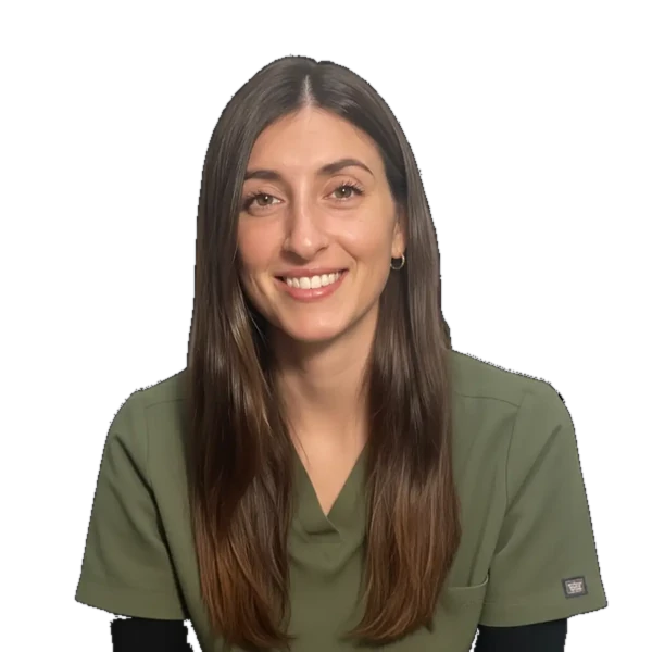
With an approach that combines Softness, Transparency and Great Meticulousness, Carolane ensures that each patient immediately feels confident and safe.
Its objective is simple: to make your care experience as comfortable as it is effective.
With a rich nursing experience that began in 2014, Carolane has enriched her expertise with a background in Medical Aesthetic Treatments To offer you the most recent protocols.
Passionate about improving the quality of life, she is entirely dedicated to supporting you with listening and professionalism throughout your career at the CARE clinic.