An often rough and brownish plaque. Its appearance near the eye justifies a clinical examination.
.png)
Seborrheic keratosis is a non-cancerous skin growth. It forms from surface cells and creates a patch that is often brownish or beige.
On the eyelid, this waxy lesion gives the impression of being stuck on. Its presence near the edge of the lashes or the surface of the eye requires special attention.
A clinical examination helps confirm its typical appearance. The doctor ensures that it does not interfere with blinking and rules out other skin conditions.
THE CARE EXPERIENCE
Signs to observe
The characteristics that often prompt a consultation.
01
Stuck-on and pigmented lesion.
02
Irregular surface to the touch.
03
Lesion near the eye.
Discomfort to the touch.
Discomfort when applying makeup or cleaning the eyelid.
Surface sensitivity.
Pigmentation to be checked.
Change near the hairs.
The approach depends on depth, lash-line position and what the doctor sees under magnification.
A stable plaque may be watched; an irritated, bleeding or lash-line lesion may need a different plan.
Schedule an assessment"
"During the examination under magnification, I verify that the patch remains strictly superficial and that it does not invade the root of your lashes or the small glands located on the edge of the eyelid."
Dr. Karen Dzolang, family doctor
Variations in appearance
The appearance guides the medical evaluation.
Observation
Differentiating the lesions
Distinct visual markers.
01
Typical
The most common form has clear edges, a stuck-on appearance, and very slow progression without bothering the eye.
Signs
•
Clear edge
•
Slow progression
•
Intact lashes
02
Irritated
Regular rubbing can make the patch red, sensitive, or covered with a small temporary crust.
Signs
•
Local discomfort
•
Friction crust
•
Provoked bleeding
03
Pigmented
Some patches accumulate more pigment and become very brown or black, requiring a check to rule out other conditions.
Signs
•
Dark colour
•
Variable colour
•
Borders to check
04
Marginal
A growth located on the free edge of the eyelid requires great caution to protect the surface of the eye.
Signs
•
Proximity to the eye
•
Eyelash deviation
•
Discomfort when blinking
Background
01
Surface cells form a visible lesion, often with a raised surface or a different texture.
02
Color, elevation, contours, thickness, rate of evolution, involvement of the eyelash margin — each detail points to a typical lesion or a sign that requires further investigation.
03
Surveillance, local excision, or pathological analysis: the decision follows what the clinical assessment has shown.
The changes that warrant an examination.
A lesion that visibly grows in a few weeks, whereas a keratosis usually develops over months or years.
Attention
Blood appearing without scratching the lesion, or bleeding that recurs with the slightest touch.
Attention
A scab that reappears after healing, or a sore that doesn't close — typical of early-stage skin cancers.
Attention
A dull ache, tenderness to the touch, or unusual burning around the plate.
Attention
A very irregular color or asymmetrical borders warrant closer attention.
Attention

Family doctor and graduate of the Université de Montréal, in good standing with the Collège des médecins du Québec (CMQ), Dr. Karen Dzolang serves as medical director for the CARE network. For eyelid lesions and skin plaques near the eye, she helps frame the evaluation, possible indications, treatment limits and situations where another medical opinion is preferable.
A registered nurse trained in pediatrics at Sainte-Justine Hospital, Karine Charbonneau later specialized in the dry eye clinic. Recognized by her patients for her gentleness, patience, and attentiveness, she supports each individual with precision and care, from the first appointment to long-term follow-up.

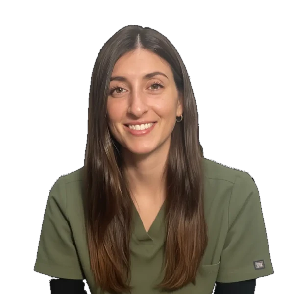
With an approach that combines Softness, Transparency and Great Meticulousness, Carolane ensures that each patient immediately feels confident and safe.
Its objective is simple: to make your care experience as comfortable as it is effective.
With a rich nursing experience that began in 2014, Carolane has enriched her expertise with a background in Medical Aesthetic Treatments To offer you the most recent protocols.
Passionate about improving the quality of life, she is entirely dedicated to supporting you with listening and professionalism throughout your career at the CARE clinic.