Surgical excision of large eyelid lesions in Greater Montreal

At Experience Care, Dr. Karen Dzolang first examines your lesion and confirms it is benign. She then explains whether excision with pathological analysis is suitable for your situation.
Le principe

How excision removes the complete lesion with laboratory analysis

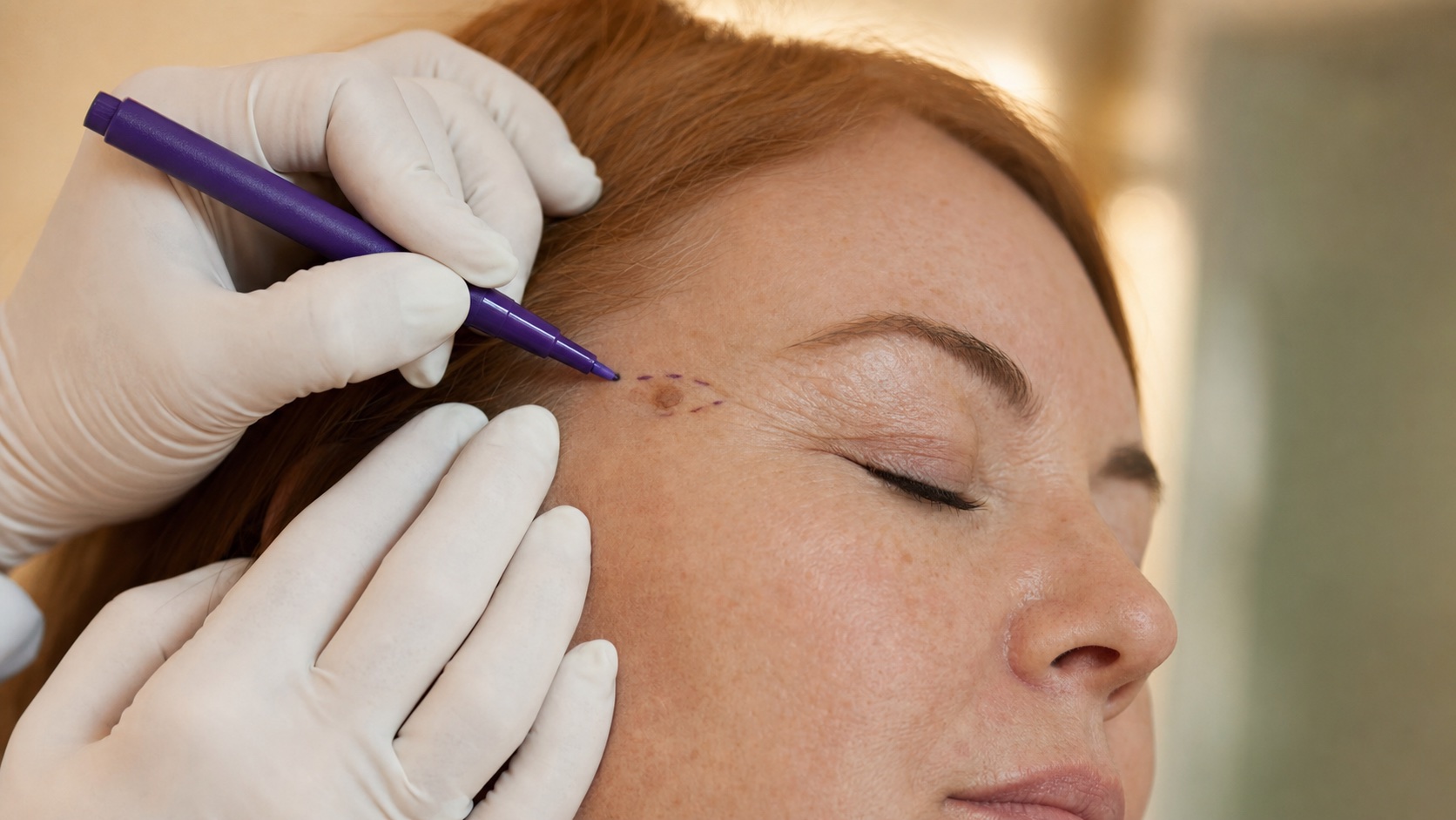
An incision along the skin's tension lines, closed with fine sutures, with the specimen sent to the pathology lab.

01

The Principle

Elliptical excision along natural tension lines, closed with fine sutures. Scar as discreet as possible.

02

The Differentiator

Every excised specimen is sent for histological analysis. It is the only modality (along with Mohs) that confirms the exact nature of the lesion.

03

Scarring

Incision follows tension lines. Two-layer closure distributes tension. Scar matures at 1 year, usually discreet.

What situations often call for excision?

Four periocular situations that Dr. Dzolang carefully assesses before recommending excision.

Which lesions can be treated with this technique?

Five periocular conditions that Dr. Dzolang treats with excision after confirming the lesion is benign and not requiring referral.

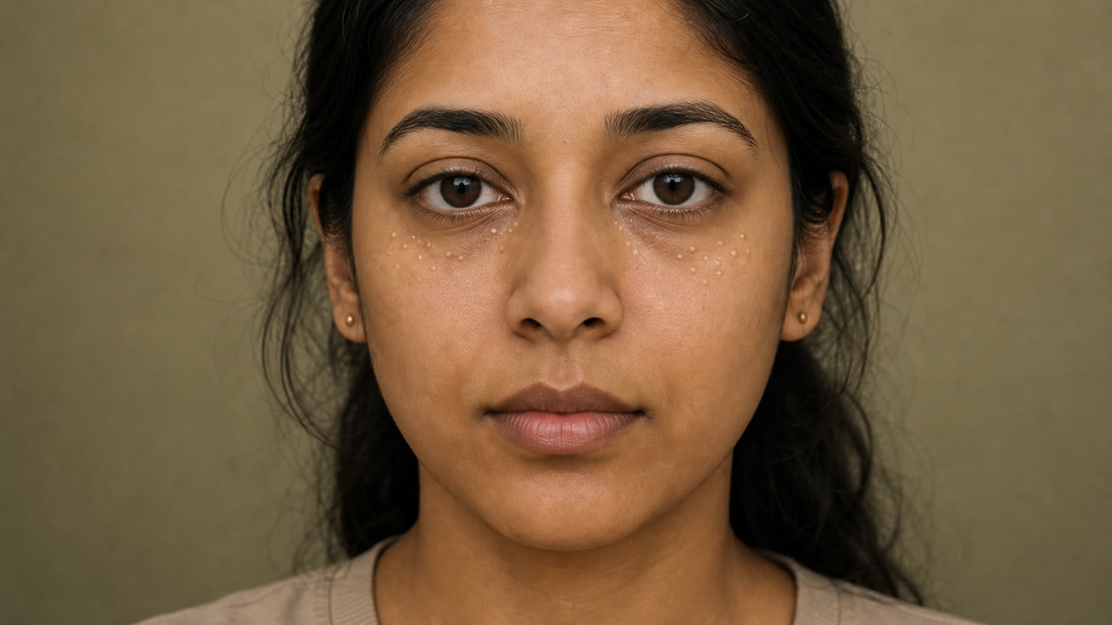
Large or atypical mole on the eyelid that changes in appearance or requires pathological analysis.

Large or recurrent xanthelasma that cannot be fully removed with other techniques (laser, plasma).

Epidermoid or sebaceous cyst on the eyelid that reoccurs if the capsule is not completely removed.

Very thick or recurrent seborrheic keratosis after initial treatment with cryotherapy or plasma.

Large pedunculated skin tag (acrochordon) that rubs or bleeds, and for which removal with sutures offers a cleaner result than cryotherapy alone.

When is excision not recommended?

Three situations where excision is referred elsewhere, and three situations where waiting or an alternative approach is preferred.

Lesion on the free eyelid margin, near the lacrimal punctum or tear ducts — mandatory referral to oculoplastics.

Suspected skin cancer (carcinoma, melanoma) on the eyelid — referral to dermatology or Mohs surgery before any removal.

Active skin infection at the excision site — such as cellulitis, stye, or open wound.

Anticoagulant medication use or uncontrolled bleeding disorder — higher risk of bleeding or hematoma.

Keloid or known hypertrophic scarring tendency.

How an in-office excision is performed

A visit lasting about 30 to 45 minutes, with a follow-up appointment for suture removal in 5 to 7 days.

01

Assessment, Marking, and Local Anesthesia

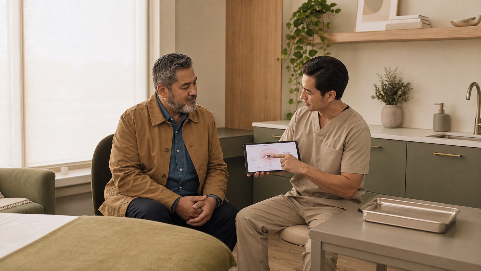
Lesion examination, marking before infiltration, local lidocaine injection. Numbing in a few minutes.

02

Elliptical excision and suture closure

Elliptical excision along natural tension lines. Specimen sent for pathological analysis. Closure with fine 6-0 or 7-0 sutures.

03

Suture removal and pathology analysis

Dressing, antibiotic ointment 5-7 days, SPF 50+ 6 months. Suture removal at 5-7 days on eyelid. Pathology analysis result provided at removal visit.

THE CARE EXPERIENCE
Beyond treatments, we offer care rooted in listening and empathy. Each patient is unique, and our greatest satisfaction is restoring the comfort you thought you had lost.
An approach appreciated by our patients. Rated 5.0/5 on Google.

Who performs surgical excision at CARE Clinic?

Dre Karen Dzolang — Directrice médicale et médecin de famille du réseau CARE
Dr. Karen Dzolang
Medical Director | Family Physician

Family Physician Trained at the Université de Montréal and a current member in good standing of the Collège des médecins du Québec (CMQ), Dr. Karen Dzolang serves as the medical director of the CARE network. For eyelid lesions like xanthelasma, she helps frame the assessment, possible indications, limits of removal, and situations where another medical opinion might be preferable.

FAQS
What happens during a surgical excision?

The doctor will first examine the lesion and mark its outline before administering a local lidocaine anesthetic. The lesion is then removed in an elliptical sample with a scalpel. The wound is closed with fine sutures. The removed tissue is always sent for pathological analysis. The procedure typically takes 30 to 45 minutes.

Is it painful? Is anesthesia used?

Local anesthesia makes the procedure painless. You may feel the initial injection, but nothing during the excision itself. Mild sensitivity afterward is normal and usually managed with a simple pain reliever.

How many visits are required?

Typically, two visits are needed: one for the excision itself, and another to remove the stitches. A third visit for scar assessment may be scheduled a few weeks later, based on your progress.

When are the stitches removed?

On the eyelid, stitches are removed after 5 to 7 days. Leaving them in longer than 7 days increases the risk of permanent marks. For thicker periocular areas, stitches are removed after 7 to 10 days.

Why is a routine pathological analysis performed?

Surgical excision is one of the only techniques that yields an intact tissue sample for histological analysis. Every excised lesion is sent to the lab to confirm its exact nature. This is especially important for large or atypical lesions and helps to rule out precancerous changes. Other techniques (cryotherapy, plasma, laser) destroy or thermally alter the lesion, making lab analysis impossible.