Surgical radiofrequency for precise eyelid lesion removal in Greater Montreal

At CARE Clinic, the physician first examines your lesion to confirm it is benign, checks for the absence of a pacemaker, and then reviews your options with you.
The principle

How Radiofrequency Simultaneously Cuts and Coagulates

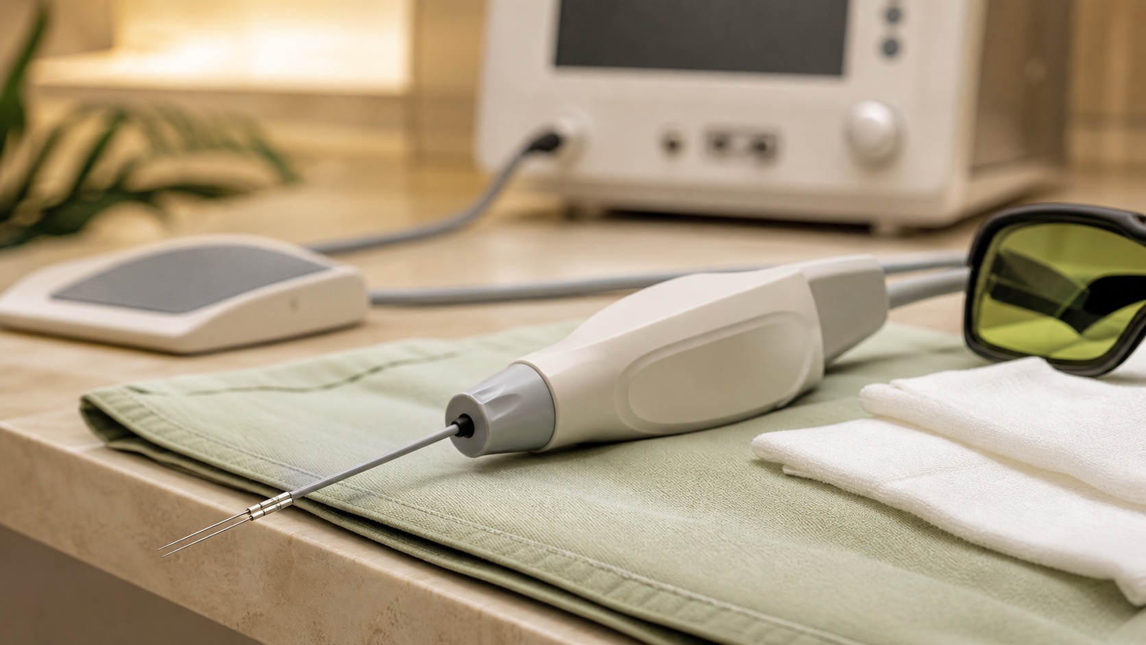
A 4 MHz current that cuts tissue and seals small blood vessels in a single step.

01

How it works

4 MHz radiofrequency cuts the lesion and simultaneously coagulates small vessels. Clean removal with very little bleeding vs scalpel.

02

Precision

Multiple modes (pure cut, blend, coagulation) adaptable. Loop or fine needle chosen based on the lesion. Surrounding healthy tissue preserved.

03

Healing

4 MHz frequency limits lateral thermal injury vs traditional electrocautery. Clean healing and scar generally discreet.

What situations often lead to considering radiofrequency?

Four periocular scenarios the physician reviews before recommending radiofrequency.

Which Lesions Can Be Treated with This Technique?

Six types of benign raised lesions the physician can remove in clinic after confirming they are benign.

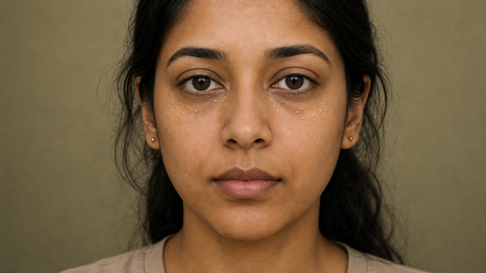
Skin growths (skin tags) on the eyelids that become bothersome or inflamed due to eyeglass friction.

Yellowish patches (xanthelasmas) on the eyelids that are not reduced by creams or dietary changes.

Thick brown or black rough patches (seborrheic keratoses) that rub against the lashes or frames of eyeglasses.

Yellowish bumps (sebaceous hyperplasia) around the eyes that increase in size and become visible.

Raised mole (dermal nevus) on the eyelid, stable and benign, that rubs against the eyelids or glasses.

When Is Radiofrequency Not Recommended?

Three situations where radiofrequency is not used, and three where another method is preferred.

Pacemaker, defibrillator, or other implanted electronic device — risk of electromagnetic interference from radiofrequency.

Active infection in the treatment area — including stye, skin herpes, or open wound.

Lesion that changes rapidly, bleeds, or has irregular borders — referral to dermatologist before any removal.

Dark skin (skin types IV to VI) — higher risk of post-inflammatory hyperpigmentation.

Anticoagulant medication use or known bleeding disorder — greater risk of bleeding during the procedure.

What Happens During a Radiofrequency Treatment Session?

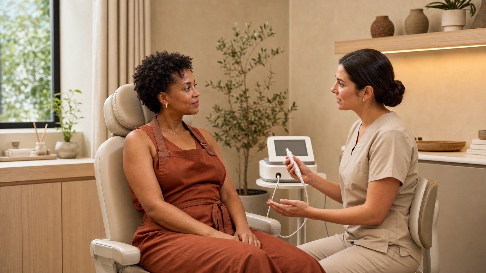
A short appointment: examination, local anesthesia, simultaneous cutting and coagulation, and home care instructions.

01

Assessment, pacemaker check, and anesthesia

Lesion examination and confirmation that it is benign. Check for pacemaker absence. Anesthetic cream 30-45 min prior and local lidocaine injection.

02

Simultaneous cutting and coagulation

A 4 MHz loop or fine needle that simultaneously cuts and coagulates. Actual procedure 1 to 3 min per lesion. The eyelid margin is avoided.

03

Healing and return to activities

You can leave immediately. Scab falls off naturally within 5 to 10 days. No makeup for 7 days. Antibiotic ointment for 5 to 7 days. SPF 50+ for 8 weeks.

THE CARE EXPERIENCE
Beyond treatments, we offer care rooted in listening and empathy. Each patient is unique, and our greatest satisfaction is restoring the comfort you thought you had lost.
An approach appreciated by our patients. Rated 5.0/5 on Google.

Who Performs Radiofrequency at CARE Clinic

Dre Karen Dzolang — Directrice médicale et médecin de famille du réseau CARE
Dr. Karen Dzolang
Medical Director | Family Doctor

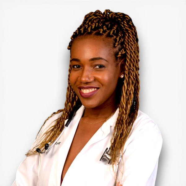
Family doctor and graduate of the Université de Montréal, in good standing with the Collège des médecins du Québec (CMQ), Dr. Karen Dzolang serves as medical director for the CARE network. For eyelid lesions and skin plaques near the eye, she helps frame the evaluation, possible indications, treatment limits and situations where another medical opinion is preferable.

FAQS
What happens during a surgical radiofrequency session?

The practitioner (physician or optometrist, depending on the lesion) will first examine the lesion to confirm it is benign and check for any implanted electronic devices (such as a pacemaker or defibrillator). A numbing cream is applied 30 to 45 minutes before the procedure, followed by a lidocaine injection. The 4 MHz radiofrequency device cuts and coagulates the lesion simultaneously. The session lasts about 15 to 30 minutes per lesion.

Is the procedure painful? Will anesthesia be used?

Local anesthesia (numbing cream and lidocaine injection) ensures the procedure is comfortable. You might feel some mild pressure, but there shouldn’t be pain. It is normal to have some sensitivity for a few hours afterwards, which typically resolves within 24 to 48 hours.

How many sessions are needed?

Most lesions— such as skin tags, milia, sebaceous hyperplasia, and seborrheic keratoses— typically require only one session. Larger xanthelasmas may need a second session a few weeks later. The practitioner will determine the treatment plan after assessing your lesion.

How long does healing take?

A scab will form on the treated area and usually falls off naturally within five to ten days. Any lingering redness typically fades in two to four weeks. Final skin pigmentation generally stabilizes around two to three months. To help prevent hyperpigmentation, sunscreen with SPF 50+ is recommended for eight weeks after the procedure.

Why is having a pacemaker an absolute contraindication?

Radiofrequency emits electromagnetic signals that can interfere with a pacemaker or an implanted defibrillator— potentially causing interruption, reset, or inappropriate shocks. On the eyelid, the proximity to the chest makes this interference especially risky. For patients with implanted electronic devices, other techniques such as cryotherapy or JETT Plasma are recommended.